Leading Tech

PET-CT

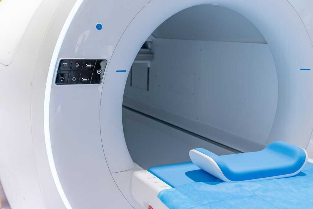
A PET-CT scan (Positron Emission Tomography–Computed Tomography) is an advanced imaging technique that merges two powerful technologies—PET, which reveals how your body’s cells and tissues are functioning by capturing metabolic activity, and CT, which provides detailed anatomical images of organs, bones, and tissues. During the procedure, a small amount of radioactive tracer (usually a form of glucose called FDG) is injected into the body, and as cancer cells and other active tissues absorb more of this tracer, the scanner detects these areas of high activity; the combined PET-CT images then allow doctors to accurately identify the location, size, and behavior of diseases, aiding in precise diagnosis, treatment planning, and monitoring of various conditions.

Read More

4D CT

A 4D CT scan is an advanced imaging technology that captures detailed three-dimensional images of the body along with the added dimension of time, showing motion such as breathing or organ movement in real time. This dynamic imaging helps doctors accurately plan radiation therapy by visualizing how tumors and surrounding organs move, ensuring precise targeting and minimizing radiation exposure to healthy tissues. It is especially useful in treating cancers of the chest and abdomen, where organ movement can affect the accuracy of treatment.

Read More

DEXA Scan

A DEXA scan (Dual-Energy X-ray Absorptiometry) is an advanced and highly precise imaging test that measures bone mineral density (BMD) to assess bone strength and detect conditions like osteoporosis and osteopenia. Using two low-dose X-ray beams, the scan provides detailed images of areas such as the spine, hips, or sometimes the whole body, allowing doctors to evaluate bone loss and fracture risk with great accuracy. The procedure is quick, non-invasive, and painless, typically completed within minutes, and is widely recommended for postmenopausal women, older adults, or individuals at risk of bone diseases. Regular DEXA scans help monitor changes in bone density over time, guiding effective prevention and treatment plans for maintaining long-term bone health.

Read More

Brachytherapy

A Brachytherapy is an advanced form of internal radiation therapy used to treat cancer by placing a small radioactive source directly inside or near the tumor. This targeted technique delivers a high dose of radiation precisely to the cancer cells while minimizing exposure to surrounding healthy tissues, allowing for effective treatment with fewer side effects. It is commonly used for cancers of the cervix, prostate, breast, and other localized areas, and can be performed as a temporary or permanent implant depending on the type and stage of the disease.

Read More

TrueBeam Linear Accelerator (LINAC) Radiation Therapy

The TrueBeam Linear Accelerator (LINAC) Radiation Therapy is a cutting-edge cancer treatment technology designed to deliver precise, high-energy radiation directly to tumors while minimizing exposure to surrounding healthy tissues. It uses advanced imaging and motion management systems to track and target tumors with exceptional accuracy, even as the patient breathes or moves slightly. TrueBeam can perform a wide range of treatments—including IMRT (Intensity-Modulated Radiation Therapy), IGRT (Image-Guided Radiation Therapy), SRS (Stereotactic Radiosurgery), and SBRT (Stereotactic Body Radiation Therapy)—all with speed, safety, and comfort. This technology enables doctors to treat complex cancers of the brain, lungs, breast, head and neck, and other areas with greater precision and fewer side effects, resulting in more effective and patient-friendly cancer care.

Read More

Mammography

A Mammography is a specialized medical imaging technique that uses low-dose X-rays to create detailed images of the breast, helping in the early detection and diagnosis of breast diseases, especially breast cancer. It can identify tiny lumps or abnormalities that are often too small to be felt during a physical examination. The procedure is quick and safe, involving gentle compression of the breast to capture clear images from different angles for accurate evaluation. Screening mammograms are recommended for routine breast health checks, while diagnostic mammograms are used to investigate specific symptoms or findings, making mammography one of the most effective tools for saving lives through early detection.

Read More

Magnetic Resonance Imaging (MRI)

A Magnetic Resonance Imaging (MRI) scan is a non-invasive diagnostic test that uses strong magnetic fields and radio waves to create highly detailed images of the body’s internal organs, tissues, and structures. It provides clear and accurate visualization of areas such as the brain, spine, joints, muscles, and organs without using any radiation. MRI is especially useful for detecting tumors, brain and spinal cord conditions, joint injuries, and soft tissue abnormalities. No ionizing radiation is produced during an MRI exam, unlike X-rays. The procedure is safe and painless, offering doctors precise information to diagnose conditions and plan effective treatments with confidence.

Read More
Chat With Us